- 65 Mario Capecchi Drive, Salt Lake City, Utah,84132
- 175N 400W, #C10, Orem, Utah,84057
- 2255N 1700W, Layton, Utah,84041
- 1054 E. Riverside Drive, St. George, Utah,84790
- 552 N. Dixie Drive, St. George, Utah,84770
- Harley Street, London, ,W1G8
- 1025 E. 3300 S., Suite B, Salt Lake City, Utah,84106

Orbital Tumors - Hemangiopericytoma
Description of Orbital Tumors - Hemangiopericytoma
- Contractile cells that wrap around {the} endothelial cells of blood vessels are known as pericytes. Orbital hemangiopericytoma is a rare, solid, slow-growing tumour that arises from {the} proliferation of pericytes in {the} orbital blood vessels but can involve blood vessels in {the} conjunctiva, choroid, optic nerve or skin of medial canthus.
- Hemangiopericytoma constitutes 1-3% of all biopsied lesions of {the} orbit and 1% of all lacrimal sac tumours. This tumour might be benign or malignant, and starts around 40 years of age with a predilection for men.
Pathophysiology of Orbital Tumors - Hemangiopericytoma
- Orbital hemangiopericytoma is caused by inordinate layering of sheets of pericytes around improperly formed blood vessels within {the} orbital structures. The tumour cells contain few cytoplasmic organelles and are formed from pluripotent mesenchymal cells surrounding {the} blood vessels. “Staghorn” vessels are thin-walled, branching blood vessels seen specifically in hemangiopericytomas.
- They have ovoid or spindle-shaped nuclei. 75% of {the} lesions are encapsulated and well-circumscribed while 30% of orbital hemangiopericytomas look very similar to a malignancy. They stain uniformly for CD34 and vimentin while 70% are positive for Leu-7.
Symptoms and Signs of Orbital Tumors - Hemangiopericytoma
- Clinical presentation is variable. Patients might present with any of {the} following symptoms:
- Painful or painless, slow-growing mass in {the} orbit
- Proptosis and exophthalmos
- Raised pressure within {the} orbit
- A painless mass near {the} medial canthus might be a lacrimal sac hemangiopericytoma
- Epiphora or watering of {the} eyes
Orbital hemangiopericytoma commonly involves {the} superior orbit and are commonly ballottable on palpation. There might be visual loss, proptosis during exophthalmometry and decreased extraocular muscle motility depending on location of {the} tumour.
Diagnosis of Orbital Tumors - Hemangiopericytoma
- Even though proper clinical examination will aid in reaching a probable diagnosis, imaging studies like Computed Tomography or Magnetic Resonance Imaging help in planning for surgical excision. Definitive diagnosis is reached only through histopathologic evaluation of {the} tumour.
- Hemangiopericytomas are easily confused with various orbital masses like fibrous histiocytoma, hemangioma, glomus tumour, sarcoma and vascular malformation.
Treatment of Orbital Tumors - Hemangiopericytoma
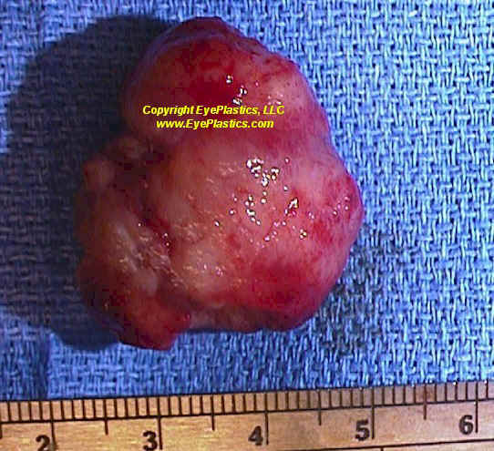
- Definitive management of a case of hemangiopericytoma is complete local excision of {the} tumour along with {the} capsule. Maintaining proper haemostasis during surgery is of paramount importance as {the} tumour is highly vascular.
- There has been some buzz about {the} role of chemotherapy and radiotherapy in preoperative management of {the} tumour, but with limited benefit.
- There is an overall 89% 5-year-survival rate seen in hemangiopericytoma. There is also a possibility for local recurrence and local metastasis, but distant metastasis to lung, liver, bone and mediastinum is a rare occurrence.