

Procedures
- Home
- Anophthalmos
- Blepharoplasty
- Botulinum
- Brow Lift
- Congenital
- Dry Eye
- Eyelid Laxity
- Infections
- Inflammation
- Lacrimal System
- Lagophthalmos
- Latisse
- Mid Face Lift
- Orbital Tumors
- Ptosis
- Skin Rejuvenation
- Skin Tumors
- Symblepharon
- Thyroid
- Trauma
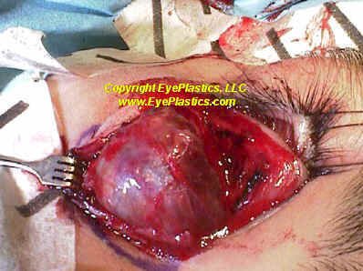
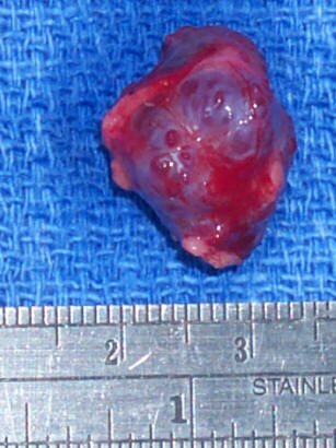
Cavernous Hemangioma
General
- most common benign solitary lesion in adults
- 30-60 year old female is typical
- slow axial proptosis over 3-5 yrs, averaging 5mm
- retinal striae
- hyperopia
- strabismus
- Optic nerve compression
- rapid growth in pregnancy
- increased intraocular pressure
Imaging
- CT: smooth discrete lesion, fills with dye after 20 min; coronal cuts important to know tumor position relative to optic nerve. for sugical plan
- MRI: hypointense to fat on T1, hyperintense to fat on T2
- U/S: high reflectivity (A-scan high amplitude internal echoes)
Pathology
- well encapsulated and tolerated
- shows large cavernous spaces with red blood cells
Surgery
- surgery for symptoms especially optic nerve compression
- usually lateral orbitotomy with complete resection from intraconal position generally feasible


