

Ocular Implants
Implant Typesmagnets, gold, silver, glass, silicone, cartilage, bone, fat, cork, titanium mesh, acrylics, wool, rubber, catgut, peat, agar, polyethylene, hydroxyapatite |
 |
MEDPOR® Biomaterial
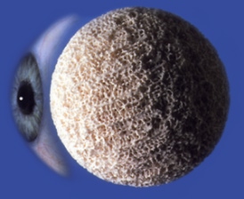
MEDPOR® is comprised of a lightweight, porous form of high-density polyethylene, a material with a long history of medical applications. Its unique, highly porous texture allows vessels to incorporate into the enhancement shape, integrating MEDPOR® into a patient's tissues. The shape and size can be customized by your surgeon to fit your individual needs. MEDPOR® eliminates the need for grafts or silicone implants.
|
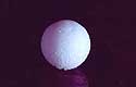
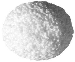
MEDPOR® Orbital Spheres Surgeons may select from sphere implant diameters of 14 mm to 22 mm. A resterilizable sizer set is available for selecting the appropriate implant diameter at the time of surgery. |
COI ®Implants
The Conical Orbital Implant (COI) ® was designed and developed to address many of the common problems associated with the correction of the anophthalmic socket. The COI® design provides removal of the anterior surface of the channels in the medial, lateral and inferior quadrants. |
Bio-Ceramic
|
Bio-Ceramic:
|
 |
|
Hydroxyapatite:
|
 |
Surgical Choices and Techniques
- In 1989, corraline sphere shaped implants were introduced. Hydroxyapatite is an inert, biocompatible and nontoxic material that has been in use in the medical field for over 15 years. Hydroxyapatite is a calcium phosphate hydroxide compound made up of multiple interconnecting pores. Because this is an inert porous substance, once implanted into the orbit it becomes vascularized and hence an integral part of the orbit. In recent years, porous polyethylene implants have been utilized in a similar fashion
- Ocular Implants provide surgeons with porous biocompatible implants for orbital reconstruction following enucleation and evisceration procedures. The interconnecting, omni directional pore structure of the MEDPOR® Biomaterial allows for rapid vascularization and soft tissue in growth.
- Surgeons may select from sphere implant diameters of 14mm to 22mm. A resterilizable sizer set is available for selecting the appropriate implant diameter at the time of surgery.
- It may may be easily shaped with a scalpel. Its lightweight property, the biocompatibility of the porous polyethylene, and the ability to place the implant deep in the socket contribute to the overall popularity of this implant.
- Motility may be enhanced by suturing the extra ocular muscles directly to the implant or to an overlying fascia or scleral wrap. As with all orbital implants, it is important to have a tension free closure over the implant.
|
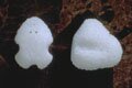
Other Shapes and Options:
|
 |
|
 |
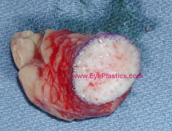
Dermis-fat graft
- is composed of subcutaneous fat and overlying dermis.
- Its advantages include the fact that it is an autologous graft and, thus, lacks concerns for bio-compatibility in disease transmission. However, there is a certain degree of fat atrophy which may occur leading to somewhat unpredictive and result of volume.
- Dermis fat graft may be used as a primary procedure following primary reconstruction for orbital exenteration, as a low cost means to provide an orbital implant and in instances in which enucleation is performed early in childhood since the fat may grow and provide stimulus for orbital growth.
- Dermis fat graft can also be used in many secondary procedures such as a replacement of an extruded orbital implant and correction of deep superior sulcus deformity.
|
 |
|
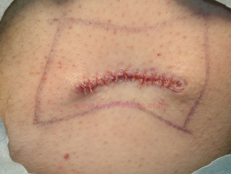 |
Complications from dermis fat graft include atrophy, central graft ulceration, granuloma formation, cyst formation, keratinization, hair growth, symblepharon, or/and donor site morbidity.


