

Canalicular Lacerations
Overview
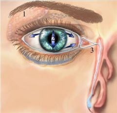 1. Lacrimal Gland 2. Tear Film on the eye 3. Canalicular and Nasolacraiml duct |
Canalicular lacerations are breaks (interruptions) in the normal tear duct drainage system. If not repaired promptly, tearing will usually result.
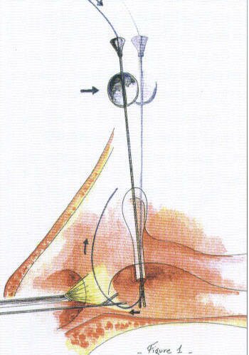
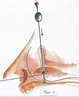
This systems originates with the puncta (there is one in both the upper and the lower eyelid) and is a conduit for tears to travel from the eyelid through the nasolacrimal sac into the nose. Tension, from trauma such as a blow from the fist, can result in an eyelid laceration which involves the canalicular system. Repair requires re-approximation of the eyelid as well as re-approximation of the conduit; this is best achieved with a stent such as with silastic and fine sutures such as 6,7, or 8-0 vicryls.. Download |
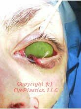
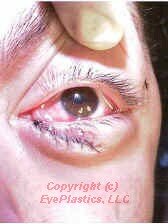
The photos below show a patient who was hit in their right eye with a fist and who sustained a canalicular laceration:
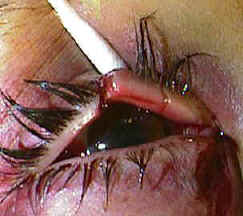 |
 |
Treatment
There are several different means to repair such an injury. Placement of a stent (silastic tubing) helps maintain proper alignment of the conduit and prevent stricture after the repair.
- Bi-canalicular stent
This places places a silicone stent in both the traumatized (lacerated) canalicular system as well as the normal. One disadvantage of this technique is the potential damage to the "good" canalicular system.
-
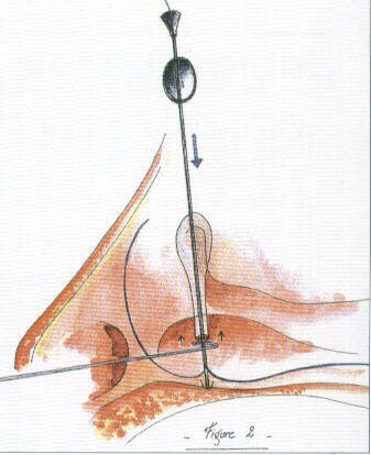
Mono-canalicular stent
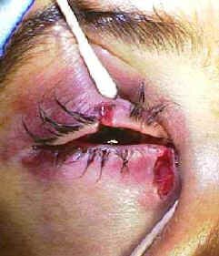
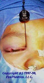
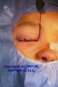
This places places a silicone stent ONLY in the traumatized (lacerated) canalicular system and thus avoids potential damage to the "good" canalicular system. A mini-Monoka or Monoka monocanalicular stent is typically usedThese three photos show a canalicular laceration and its repair with a Monoka monocanalicular stent.
 |
 |
 |
|
||
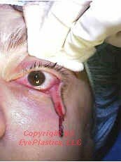
- Ritleng mono-canalicular stent
This is a type of monocanalicular stent that facilitates repair.
|
 |
 |
|
|
 |
 |
|
These two photos show a canalicular laceration and its repair with a monocanalicular stent using the Ritleng probe.
- Pig-tail probe
This allows intubation (stent placement) of the abnormal canalicular system and the normal system without entering the nose. One disadvantage of this technique is the potential damage to the "good" canalicular system. A Goldberg Bicanalicular Cerlage is used for stenting the upper canalicular system system for reconstruction, truama, and chronic stensosis of the upper system.
References
- Conlon MR, Smith KO, Cadera W, Shun D,Allen LH. An animal model studying construction techniques and histopathologic changes in repair of canalicular a lacerations. Can J Ophthalmol 1994;29:3d
- .Kennedy RH May J, Dailey J, Flanagan JC Canalicular laceration: an 11 year epidemiologic and clinical study. Ophthalmic Plastic and Reconstructive Surg
- Loft HJ.Wobig JL. Dailey RA. The bubble test: an atraumatic method for canalicular laceration repair. Ophthalmic Piastic ann Reconstructive Surg 1996;12:61-64.
- Long JA. A method of monocanalicular silicone intubation. Ophthalmic Surg 1988 19 204 205
- McLeish WM Bowman B, Anderson RL The pigtail probe protected by silicone intubation a combined approach to cana icu ar reconstruction. Ophthalmic Surg 1992;23:281-283.
- Reifler DM. Management of canalicular laceration. Surv Ophthalmol 199136:11 j132
- Ritleng, Peirre. A simplifed technique for lacrimal intubation. Ocular surgery news. Vol 14, No 7
// ]]>


